Autopsy report of Dubinina
city of Ivdel
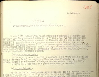
Act №4
Medical-Forensic Examination of a body
On the May 9, 1959 in accordance with decree of the Prosecutor office of Sverdlovsk region of May 7, 1959 by forensic experts of the regional forensic investigation bureau V.A. Vozrozhdenniy in the presence of the criminal prosecutor of regional prosecutor office junior Counselor in Justice L.N. Ivanov and criminal expert Churkina G.A. in the morgue of the medical unit of the PO Box 240 with daylight and sunny weather there was performed the autopsy of the body of Lyudmila Aleksandrovna Dubinina, 20 years old, in order to determine the cause of death and answers to the questions given in the declaration.
Case circumstances:
On January 23, 1959 the independent group of hikers consisting of 10 people went across the ski track Ivdel – Mount Otorten. From the 2nd Northern site the group consisted of 9 people. On February 1, 1959 the group started the climb on the mount Otorten and in the evening they put up a tent at the height of 1 079 meters.
In the night of February 2 at the unknown circumstances all 9 people died.
А.External examination:
On the section table there is a body of woman dressed as follows: head covered by a knitted cap. A worn grayish-brown wool sweater with beige wool sweater underneath, checkered shirt with buttoned sleeves. Yellow t-shirt with short sleeves, white cotton bra buttoned with three buttons. The body is wearing torn dark cotton trousers with an elastic waistband. The trousers are very torn and burned in places.
Left leg – part of the leg and foot are covered with burned grey wool cloth from a jacket with its sleeve. On the left leg there is a torn grey woolen sock. On both legs there are torn blue cotton socks with grey wool machine-knitted socks under them. Black cotton tights, torn in the crotch area, with an elastic waistband.
On the legs of the body there are light brown cotton stockings. The stocking from the left leg is removed, the right stocking is held with an elastic band. Grey ladies belt with elastic supports. Satin briefs. The belt is buttoned with black buttons.
After the removal of the clothes there was found: a female body of proper constitution and good nutrition, 167 cm long. The post mortem lividity is of bluish grey color, particularly on the posterior and lateral surface of neck, torso and extremities. Blond hair on the head, braided into one braid up to 50 cm long with a blue silk ribbon. The forehead is straight, retreating to the back.
The skin of the face is of yellowish brown color. The soft tissue in the area of the supraorbital ridges, on the bridge of the nose, the eye sockets and left temporal-zygomatic area are all absent, exposing the facial skull bones. The eye sockets are glaring, the eyeballs are absent. The bones of the nasal bridge are intact, the nasal cartilage is flattened. There is an absence of the soft tissue from the upper lip on the right with thinning of its edges, exposing the alveolar edge of the upper jaw and teeth. The teeth are regular, intact. The tongue in the oral cavity is absent. The oral mucosa are of grayish green color.
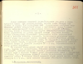
- 2 -
The ear pinnae are oval. The orifices of mouth, nose and ear passages are clean. The neck is long and thin. The soft tissue in the neck area is flaccid when palpated. When palpating the neck, there is extraordinary mobility of the thyrohyal and thyroid cartilages. The chest is of cylindrical shape. The breasts are of medium size, full. The nipples and areoles are of pale brown color. The stomach is located at normal chest level. The external genitals are formed properly. The hymen is of ring-like shape with high rim, meaty. The natural orifice of the hymen allows the passage of adult’s pinkie tip. The mucosa of the vagina is of purple-red color. On the external and anterior surface of the left thigh in the middle third there is diffuse bruising of bluish-purple color of 10 x 5 cm with deep skin hemorrhage.
On the back of the hand there is soft tissue that is dense upon palpation, the fingers of the hands are semi-bent. The end phalanges of the fingers are covered with wrinkled ‘bath skin’ which is removed together with the nail plate. In the area of the feet and fingers the ‘bath skin’ is of pale grey color with purple shade. During palpation of the chest there is noted an extraordinary mobility of the ribs. In the area of the left temporal bone there is a soft tissue defect sized 4 x 4 cm with the bottom of the defect exposing the temporal bone.
B.Internal examination.
The skin pieces of the scalp from the internal surface are wet, juicy and shiny. The bones of the skull vault and base are intact. The brain membrane is bluish from poor blood filling. The cerebral gyri convolutions are poorly defined. The grey brain matter is poorly differentiated from the white. The contours of the lateral brain ventricles are poorly defined. The vessels of the brain base are without particularity. The subcutaneous fatty tissue of the trunk is well developed. The position of the internal organs is regular, the pleural cavities contain up to one and a half litres of liquid dark blood. The pericardium contained up to 20 cubic cm of yellowish transparent liquid. The heart is sized 12 х 4 х 5 cm. In the area of the left ventricle there is an irregularly oval-shaped hemorrhage sized 4 х 4 cm with diffuse suffusion of the right ventricle muscle. The thickness of the left ventricle muscle is 1.4 cm, the right - 0.5 cm. In the right and left halves of the heart there was up to 50 cm3 of liquid dark blood. The cardiac valves of the aorta and pulmonary artery are smooth, thin, and shiny. The coronary vessels of the heart are freely passable. The internal surface of the aorta is smooth and clean. The lungs from the surface are of bluish-red color, fluffy during palpation. A section of lung tissue is of dark red color, during pushing from the surface there is plentiful foamy bloody liquid, the lumen of the bronchi is free. The hyoid cornua are with unusual mobility (broken), soft tissue adjacent to the hyoid bone is of dirty grey color. The diaphragm of the mouth and tongue is absent. The upper edge of the hyoid bone is bared. Esophagus mucosa and bronchial tracheas are of bluish-red color. The stomach contains up to 100 cm3 of dark brown mucosal mass. The gastric mucosa is porous, of bluish-red color. The pancreas is small and lobular when sectioned and is bluish-red. The liver from the surface is smooth and opaque. The size of the liver is 23 х 12 х 10 х 6 cm.
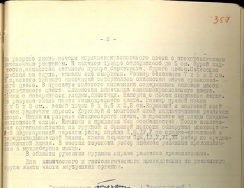
- 3 -
When sectioned the tissue of the liver is of brownish red color with a poorly defined pattern. The gall bladder contains up to 5 cm3 of brown liquid. The mucosa of the gall bladder is velvety, brown. The spleen is flabby when palpated, and the channels are still wrinkled. The spleen size is 7 х 5 х 2 cm. In the lumen of the small intestine there is mucosal mass of dirty yellow color. In the lumen of the colon there are fecal masses of dirty green color. The intestinal mucosa is of bluish red color. The surface of the kidneys is smooth and shiny. The capsule is removed easily from the kidneys. The tissue of the kidneys when sectioned is of dark red color. The size of the right kidney is 7 x 5 х 2 cm, the size of the left kidney is 9 х 5.5 х 2.3 cm. The cortex and medulla areas of the kidneys are well defined. The cortex and medulla area of the adrenals is poorly defined. The uterus upon section is pale grey, its lumen contains traces of pale yellow mucosa. The ovaries and appendages are without particularities. After the removal of the organ complex from the thoracic and abdominal cavity, the multiple bilateral fracture of the ribs was found on the right II, III, IV, V on the mid-clavicular and mid-axillary line; on the left there is a fracture of II, III, IV, V, VI, VII ribs on the mid-clavicular line. In the place of the rib fractures there are diffuse hemorrhages into the intercostal muscles.
In the area of the manubrium of the sternum on the right there is diffuse haemorrhaging.
Parts of the internal organs were taken from the body for chemical and histological study.
| Medical examiner | signature | (Vozrozhdenniy) |
| Criminal Persecutor of the Regional Persecutor Office Junior Counselor of Justice |
signature | (Ivanov) |
| Forensic expert Sverdlovsk |
(Churkina) |
Conclusion:
Based on the forensic examination of the body of L. A. Dubinina I think that the death of Dubinina was caused by massive hemorrhage into the right ventricle, multiple bilateral rib fractures, and internal bleeding into the thoracic cavity.
The said damage was probably caused by an impact of great force causing severe closed lethal trauma to the chest of Dubinina. The trauma was caused during life and is the result of high force impact with subsequent fall, throw or bruise to the chest of Dubinina.
Damage to the soft tissue of the head and ‘bath skin’ wrinkling to the extremities are the post-mortem changes (rot and decay) of Dubinina’s body, which was underwater before it was found.
Dubinina died a violent death.
Medical examiner signature (Vozrozhdenniy)


