Autopsy report of Thibeaux-Brignolle
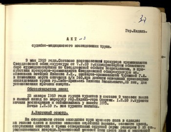
Sheet 30
city of Ivdel
Act №3
Medical-Forensic Examination of a body
On the May 9, 1959 in accordance with decree of the Prosecutor office of Sverdlovsk region of May 7, 1959 by forensic experts of the regional forensic investigation bureau V.A. Vozrozhdenniy in the presence of the criminal prosecutor of regional prosecutor office junior Counselor in Justice L.N. Ivanov and criminal expert Churkina G.A. in the morgue of the medical unit of the PO Box 240 with daylight and sunny weather there was performed the autopsy of the body of Thibeaux-Brignolle Nikolay Vasilyevich, 23 years old, in order to determine the cause of death and answers to the questions given in the declaration.
Case circumstances:
On January 23, 1959 the independent group of hikers consisting of 10 people went across the ski track Ivdel – Mount Otorten. From the 2nd Northern site the group consisted of 9 people. On February 1, 1959 the group started the climb on the mount Otorten and in the evening they put up a tent at the height of 1 079 meters.
In the night of February 2 at the unknown circumstances all 9 people died.
On the examination table is a male body clothed as follows: the head is covered by a tightly tied green woolen sports cap with three round holes sized 3 x 3 cm located in the front. A khaki canvas fur helmet with a zipper fastener [like a flying helmet]; the helmet is drawn by a cord. A green canvas sheepskin jacket with a zipper and two pockets. In the right pocket there are grey gloves. In the left pocket are 10, 20, and 2 kopeek coins; two folded pieces of paper; and a comb. A ragged wool sweater is worn on the left side. A worn-out knitted blue shirt, which on the right and bottom has torn ovals in the fabric 2 x 3 cm in size. On the left forearm there are two watches: a Sportivnye watch showing the time 8 hours, 14 minutes, 24 seconds, and a Pobeda brand watch showing the time 8 hours, 39 minutes. The legs are covered with practically new grey felt boots (valenki). On the right leg are white hand knitted wool socks; the same socks are also on the left leg. There are crumpled brown wool socks located in the soles of the corresponding felt boots. The body is wearing warm woolen winter pants, the cuffs of which are fasted by a leather belt with a metal buckle. Under these pants are blue cotton sports pants and black satin underwear. In the pocket (crossed) of the outer pants a white metal button and a metal chain from a wall clock were found.
After the removal of the clothes, the following was found: a male body of proper constitution and good nutrition, 174 cm long. The body has purple green spots on the posterolateral surface of the chest, neck and extremities. Rigor mortis has resolved in the muscle groups of the joints. The skin of the face, body and limbs is of a grey-greenish color seeping from the surface layer of the epidermis. There is hair on the head with a length of 8 cm. The forehead is high and sloping backwards and there are thick black eyebrows. The eyes are closed; the eyeballs are sunk far into their sockets. The cornea is cloudy and dry, the iris is light green, and the mucosal membrane of the eyelid is of a pale grey color. The bridge of the nose is straight.
(crossed) The bones of the nose are intact when touched. On the upper left jaw there is a defect
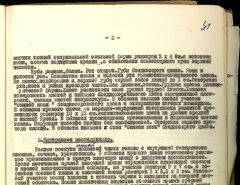
Sheet 31
- 2 -
in the soft tissue, which has an irregular oval shape with a size of 3 x 4 cm with drawn out, convoluted borders exposing the alveolar edge of the upper jaw, The teeth are white and even. The mouth is open. The lips have a pale grey color. The tongue is in the mouth. The mucous membrane of the tongue and mouth are of a dirty green color. On the cheeks, chin and upper lip there is black hair with a length of up to 1 cm. The openings of the mouth, nose and ear are clean. The neck is long and thin. The chest is cylindrical. The stomach is located below the chest. The dorsum of the hands and fingers are of a pale brown color and of parchment density; the fingers are bent at the joints. In the area of the fingers there is ‘bath skin’ of a pale grey color with the rejection of the nail plate. There is a 10 x 12 cm blue-green diffuse ecchymoma in the area of the right shoulder on the antero-internal surface at the lower middle and bottom thirds. In the area of the ecchymomare is hemorrhaging into the surrounding soft tissue. The external genitalia are normal. The opening of the anus is clear. In the area of the toes there is ‘bath skin’ of a pale grey color.
B.Internal examination.
Skin pieces of the scalp from the internal surface are juicy and dull. There is an indication of hemorrhaging from the right temple into the right temporal muscle with diffuse saturation. After dissection of the right temporal muscle determined that there was a depressed fracture in the right temporal bregmatic area on an area measuring 9 x 7 cm with a bone defect on the temporal lobe measuring 3 x 3.5 x 2 cm. This section of the bone is sunken into the cranial cavity and is located on the dura matter. After the extraction of the brain matter, in the middle cranial pit a multi-splintered fracture of the right temporal lobe was discovered with the diffusion of the cracked bone to the anterior cranial fossa and the supraorbital region of the frontal bone. A second crack runs along the front surface of the sella turcica in the area of the pterygoid processes, venturing into the interior of the underlying bone, then proceeding to the middle cranial fossa on the left with the separation of the bone from 0.1 to 0.4 cm. The dura mater, corresponding to the site of the fracture, is sharply full-blooded with the fullness of the brain substance of the right cerebral hemisphere, which differs by a more greenish-colored color. The fissures and convolutions of the brain are poorly differentiated. The brain’s grey matter is poorly differentiated from the white. The contours of the ventricles of the brain are indistinguishable. The brain is a jelly-like mass with a dirty red color. On the whole, the length of the crack in the area of the base of the skull is 17 cm. In addition, there is asymmetry due to the compression fracture of this area.
The subcutaneous tissue of the torso is sufficiently developed. The internal organs are positioned correctly. The pleural cavity is free. The pericardium contained up to 10 cm3 of red turbid liquid. The heart is 13 x 12 x 5.5 cm in size with the surface slightly covered in fat. The cardiac muscle is dark red when sectioned and soft when palpated. The thickness of the left ventricle is 1.8 cm, and the right is 0.6 cm. The right and left parts of the heart contain dark liquid blood in the form of ‘dry heart.’ The valves of heart, the aorta and the pulmonary artery are smooth, shiny, and slightly thickened. The coronary vessels of the heart are free and passable. The internal surface of the aorta is smooth and clean. The width of the aortic arch above the valves is 8.5 cm. The lungs are blue-red (blue is typed on top of another text - ed.) on the surface, creamy on touch. When sectioned the lung tissue is of a dark red color. While palpated an abundance of foamy liquid is released. The larynx and bronchus clearance is free.
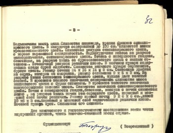
Sheet 32
- 3 -
The hyoid bone is intact. The mucosa of the esophagus, trachea and bronchi are blue-red in color. The ventricle contained up to 100 cm3 of mucous mass of a red-yellow color. The gastric mucosa has a blue-red color with good definition of the folds. When sectioned the pancreas is lobulated with a blue-red color. The surface of the liver is smooth, bright, and when sectioned its tissue has a brown-green color with hints of yellow. The hepatic pattern is poorly defined. The gall bladder contained traces of brown liquid; its mucosa was velvety brown. The size of the liver is 24 x 16 x 14 x 8 cm. The spleen is flaccid when palpated, its capsule is shriveled, and is sized 9 x 5 x 3 cm. When sectioned, the tissue of the spleen had a dark red color and gave a thick pulp when scraped.
The lumen of the small intestine contained a slimy green mass with hints of brown. Partially formed fecal matter was found in the lumen of the large intestine. The intestinal mucosa had a blue-red color. The kidneys were smooth and shiny, and the capsule from the kidney is easily removed. When sectioned the kidneys were dark red; the cortex and the medulla of the kidneys were distinguishable. The size of the right kidney is 9 x 6 x 6 cm; the size of the left is 10 x 6 x 3 cm. The cortex and the medulla of the adrenal gland were poorly distinguished. The bladder was empty and its mucous membrane was of blue color.
For chemical and histological examination, samples of the internal organs and right temporo-parietal bone were taken.
| Medical examiner | signature | (Vozrozhdenniy) |
| Criminal Persecutor of the Regional Persecutor Office Junior Counselor of Justice | (Ivanov) | |
| Forensic expert Sverdlovsk | (Churkina) |
Conclusion
On the basis of the examination of the body of Thibeaux-Brignolle, it is my opinion that his death was the result of a closed comminuted pressure fracture in the area of the base and the vault of the cranium with a prolific amount of bleeding under the meninges and brain matter while under low temperature. The above-mentioned extensive comminuted fracture of the base and the vault of the cranium are of in vivo origin and are the result of a great force with the subsequent falling, hurling and concussion of Thibeaux-Brignolle.
The corporal damage of the soft tissue in the area of the head and the ‘bath skin’ of the extremities are the result of post-mortem changes in the body of Thibeaux-Brignolle, which was found submerged in water after some time. Thibeaux-Brignolle died a violent death.
Medical examiner signature (Vozrozhdenniy)


